")
There is one other image of this object. See our image rights statement.
See more objects with the color lightgrey sienna saddlebrown saddlebrown indianred or see all the colors for this object.
Object Timeline
|
|
|
-0001 |
|
|
2013 |
|
|
2014 |
|
|
2025 |
|
Diagnostic Health-care Device, Kernel of Life (Prototype)
This is a Diagnostic Health-care Device. It was designed by Yves Béhar and manufactured by fuseproject.
This object is not part of the Cooper Hewitt's permanent collection. It was able to spend time at the museum on loan from fuseproject as part of Tools: Extending Our Reach.
In the developing world, the nearest doctor can be days away, making the diagnosis and treatment of chronic illnesses complicated. With the Kernel of Life, a medical “amulet” that tests blood, saliva, urine, and breath and transmits the results by Bluetooth to a mobile app, patients can be monitored continuously and remotely via the cloud. The superslim, two-inch (5 cm) disk slides open to reveal a microperforated pad divided into a rainbow of quadrants: red for blood, blue for saliva, yellow for urine, and green for breath. The pad separates the biofluids, parses them, and then transmits the data. With the Ker¬nel, digital medical analysis is simplified, embedded into a reusable, self-sterilizing device. Most importantly, users can monitor themselves and receive reminders about medication and treatment. The Kernel helps reduce health-care costs while expanding health coverage to underserved populations around the world, making medical care a cost-effective example of good design.
It is credited Courtesy of Gates Foundation and WIRED magazine.

- Ultrasound Machine, Vscan™
- internal: ultrasound transmitters and receivers, signal processor,....
- Courtesy of GE Healthcare.
- 13.2014.1
")
- Bioimplantable Device (England)
- polyester.
- Gift of Ellis Developments Ltd..
- 2004-15-2

- Michoacan Bone Figure Necklace
- nylon thread, bone, shell, gold.
- Judith R. Tishman Collection, gift of Peter V. and Lynn P. Tishman.
- 2002-16-2
Our curators have highlighted 2 objects that are related to this one.
, ca. 1870")
- Necklace And Pendant (possibly France), ca. 1870
- silver, enamel, jasper, pearls, glass.
- Gift of Evelyn A. Pitshke.
- 1958-64-1-a,b

- Guilloche Enamel Quatrefoil Locket Locket
- yellow gold (18 kt), white gold (18 kt), enamel, diamonds.
- Bequest of Lorey Katz.
- 2004-12-1
Its dimensions are
H x W x D (overall w/ cord): 26 × 6.7 × 1.3 cm (10 1/4 × 2 5/8 × 1/2 in.) H x W x D (amulet): 5.4 × 5.4 × 0.9 cm (2 1/8 × 2 1/8 × 3/8 in.)
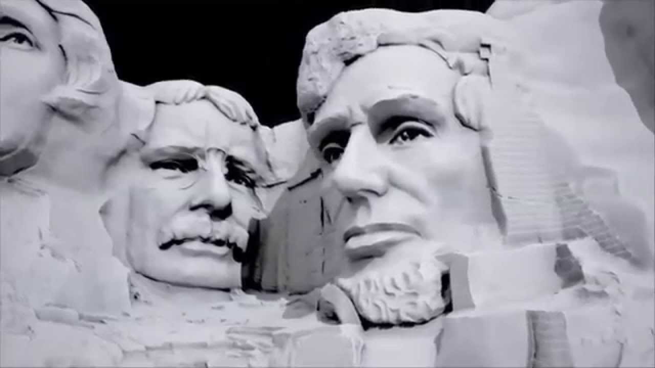

LiDAR on Mount Rushmore
A video showing how LiDAR technology was used to create a hi-res virtual model of Mount Rushmore, and how it's used in historic preservation and public education efforts.
This object was previously on display as a part of the exhibition Tools: Extending Our Reach.